Homogeneous Ana Patterns
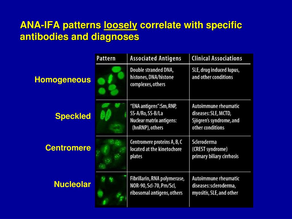
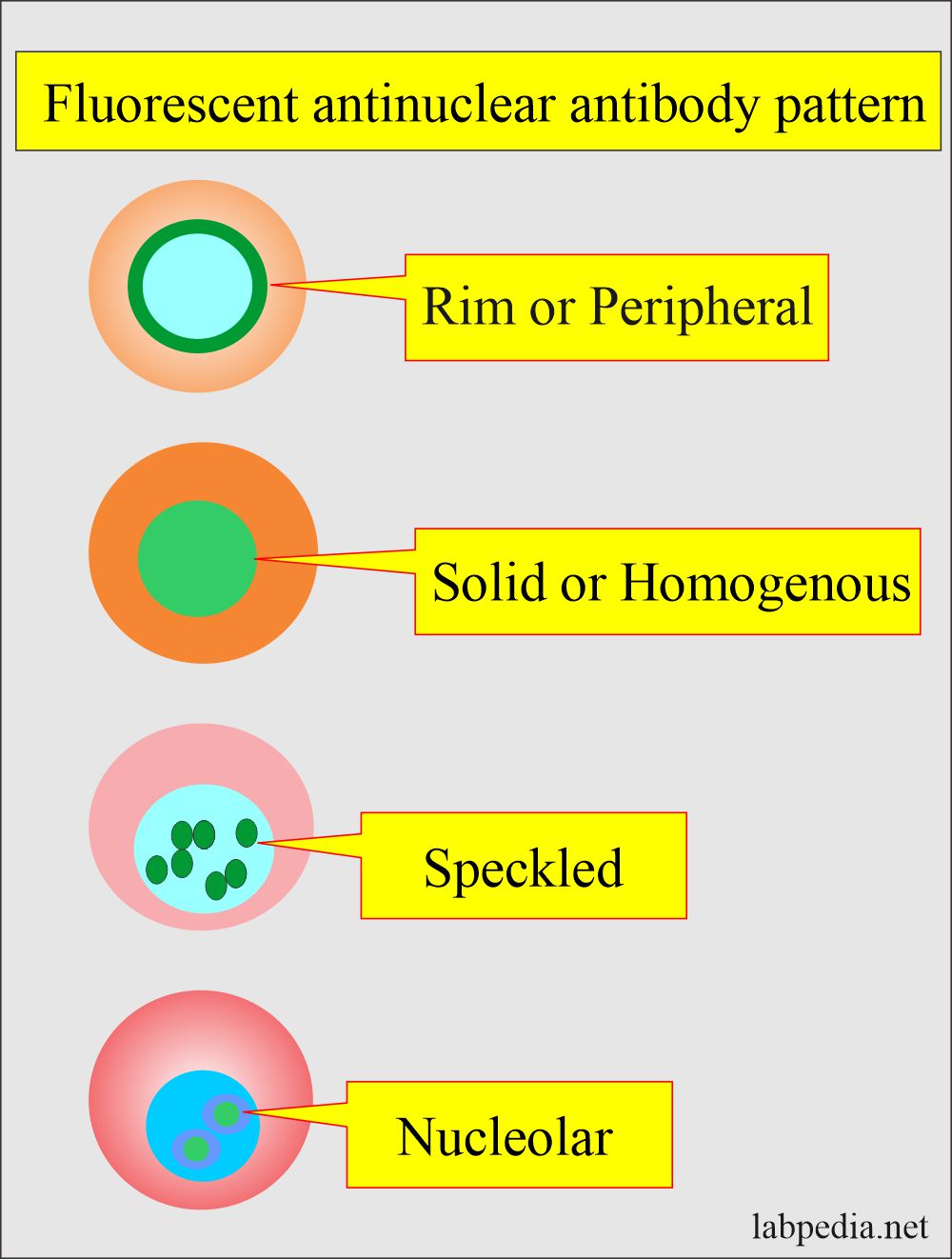
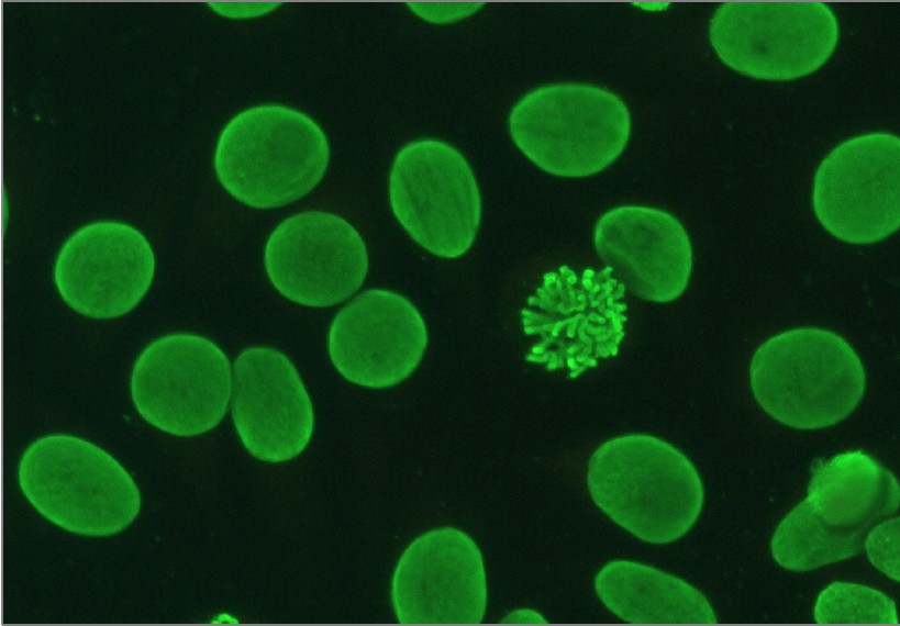
Homogeneous Ana Patterns - It’s the most common type of staining pattern. Interphase cells show homogeneous nuclear staining while mitotic cells show staining of the condensed chromosome regions. The entire nucleus is stained with ana. Talk to your provider about the meaning of your specific test results. Your immune system normally makes antibodies to help you fight infection. A titer (a measure of how much ana is in the blood) and a pattern (where the ana was detected in the cells). A homogenous pattern can mean any autoimmune disease but more specifically, lupus or sjögren’s syndrome. Total nuclear fluorescence due to an antibody directed against dna or histone proteins. Web ana patterns can be associated with different autoimmune conditions. This pattern occurs when antibodies in your blood, which usually fight infections, mistakenly target the core of your own cells. Total nuclear fluorescence due to an antibody directed against dna or histone proteins. Web each pattern is assigned an alphanumeric ac code (anticell). The commonly recognized patterns include: This is the most common pattern and can be seen with any autoimmune disease. This pattern occurs when antibodies in your blood, which usually fight infections, mistakenly target the core of your own cells. Interphase cells show homogeneous nuclear staining while mitotic cells show staining of the condensed chromosome regions. Normal value ranges may vary slightly among different laboratories. Ana stands for “antinuclear antibodies.” as. This pattern is more commonly associated with antibodies. The entire nucleus is stained with ana. What is the ana test, and why was it ordered? A titer (a measure of how much ana is in the blood) and a pattern (where the ana was detected in the cells). The commonly recognized patterns include: Web ana patterns can be associated with different autoimmune conditions. Web as they undergo treatment with steroids or other immunosuppressants, their ana. In contrast, antinuclear antibodies often attack your body's own. Homogenous staining can result from antibodies to dna and histones. Web is the ana pattern suggestive of a specific disease? The commonly recognized patterns include: Web the main ana staining patterns are homogeneous, speckled, nucleolar and centromere. Web each pattern is assigned an alphanumeric ac code (anticell). A titer (a measure of how much ana is in the blood) and a pattern (where the ana was detected in the cells). Web the presence of ana with a homogeneous & speckled (hs) pattern was significantly associated with the absence of cancer ( < 0.01). Web antinuclear antibodies (ana). Homogenous staining can result from antibodies to dna and histones. Web welcome to anapatterns.org, the official website for the international consensus on antinuclear antibody (ana) patterns (icap). A homogenous staining pattern means the entire nucleus is stained with ana. Web as they undergo treatment with steroids or other immunosuppressants, their ana pattern may become homogeneous. Ana stands for “antinuclear antibodies.”. A homogenous staining pattern means the entire nucleus is stained with ana. When active, usually a homogenous pattern on ana or less commonly speckled, rim, or nucleolar when present in high enough titer to be clinically. Web systemic lupus erythematosus (sle): Web the main ana staining patterns are homogeneous, speckled, nucleolar and centromere. Medically reviewed by carmelita swiner, md on. The entire nucleus is stained with ana. This is the most common pattern and can be seen with any autoimmune disease. Web the presence of ana with a homogeneous & speckled (hs) pattern was significantly associated with the absence of cancer ( < 0.01). Doctors may order an ana test if you have signs or symptoms of an autoimmune. Medically. Web the pattern of the ana test can give information about the type of autoimmune disease present and the appropriate treatment program. What are the most frequent causes of a positive ana? What is the ana test, and why was it ordered? In contrast, antinuclear antibodies often attack your body's own. Web welcome to anapatterns.org, the official website for the. Web patterns that are reported include, homogeneous, speckled, centromere, and others. It’s the most common type of staining pattern. The commonly recognized patterns include: When active, usually a homogenous pattern on ana or less commonly speckled, rim, or nucleolar when present in high enough titer to be clinically. Web ana patterns can be associated with different autoimmune conditions. Web antinuclear antibodies (ana) refer to an autoantibody directed at material within the nucleus of a cell. Web is the ana pattern suggestive of a specific disease? Web antinuclear antibodies (ana) represent a family of autoantibodies targeting ubiquitous cellular constituents and are a hallmark of systemic inflammatory autoimmune rheumatic diseases named connective tissue diseases (ctd). Web an ana test detects. What is the ana test? Web each pattern is assigned an alphanumeric ac code (anticell). This pattern is more commonly associated with antibodies. In contrast, antinuclear antibodies often attack your body's own. A homogenous pattern can mean any autoimmune disease but more specifically, lupus or sjögren’s syndrome. Web a homogeneous/peripheral pattern reflects antibodies to histone/dsdna/chromatin, whereas many other specificities found in systemic rheumatic diseases show speckled patterns of various sizes and densities (fine speckled, large speckled, etc.). The commonly recognized patterns include: Web patterns that are reported include, homogeneous, speckled, centromere, and others. Web ana titers and patterns can vary between laboratory testing sites due to variations in the methodology used. Web welcome to anapatterns.org, the official website for the international consensus on antinuclear antibody (ana) patterns (icap). Web systemic lupus erythematosus (sle): Web each pattern is assigned an alphanumeric ac code (anticell). Web the pattern of the ana test can give information about the type of autoimmune disease present and the appropriate treatment program. What is the ana test? Below is a summary of the patterns discussed: Many laboratories also measure pattern or the way the test looks when viewed through a microscope. This is the most common pattern and can be seen with any autoimmune disease. Fine and coarse speckles of ana staining are seen throughout the nucleus. The entire nucleus is stained with ana. What are the most frequent causes of a positive ana? Web the main ana staining patterns are homogeneous, speckled, nucleolar and centromere.
6. IFA pattern Homogeneous ANA pattern YouTube
Homogeneous Ana Pattern Pagswa

ANA Patterns
Antinuclear Factor (ANF), Antinuclear Antibody (ANA) and Its

ANA Patterns

ANA Patterns

DFS70 antibodies biomarkers for the exclusion of ANAassociated
.jpg)
ANA Mixed pattern University of Birmingham

ANA Patterns
Antinuclear antibodies (ANA) homogeneous pattern positive control
Web Antibodies That Attack Healthy Proteins Within The Cell Nucleus Are Called Antinuclear Antibodies (Anas).
Web Antinuclear Antibodies (Ana) Represent A Family Of Autoantibodies Targeting Ubiquitous Cellular Constituents And Are A Hallmark Of Systemic Inflammatory Autoimmune Rheumatic Diseases Named Connective Tissue Diseases (Ctd).
It’s The Most Common Type Of Staining Pattern.
This Pattern Is More Commonly Associated With Antibodies.
Related Post: